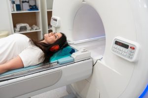
Heart failure is widespread. In Europe, more than 10 million people suffer from heart failure. For various reasons, the heart can no longer generate a sufficiently high cardiac output to supply the body with enough oxygen and enable a normal metabolism. This can be due to both reduced pumping power (systolic heart failure) and impaired relaxation capacity (diastolic heart failure) of the heart muscle. An important new study has improved the detection of heart failure in women – meaning that more female patients can be diagnosed at an earlier stage.
Magnetic Resonance Imaging to Better Diagnose Heart Failure
Researchers led by teams from the Universities of East Anglia (UEA), Sheffield and Leeds have been able to fine-tune the way magnetic resonance imaging (MRI) is used to detect heart failure in women’s hearts, making it more accurate. By refining the method specifically for women, the researchers were able to diagnose 16.5 percent more women with heart failure. This could have a huge impact on the NHS (National Health Service in the UK), which diagnoses around 200,000 patients with heart failure each year, according to the study’s lead author, Dr. Pankaj Garg from the University of East Anglia’s Norwich Medical School and consultant cardiologist at Norfolk and Norwich University Hospital. This improved method will improve early detection, meaning more women can receive life-saving treatment sooner. In 2022, the UEA and the University of Sheffield published research showing how MRI scans can be used to detect heart failure, leading to the technique being widely used by medical professionals.

When a heart begins to fail, it is no longer able to pump blood out effectively, so the pressure inside the heart rises. Currently, one of the best ways to diagnose heart failure is to measure the pressure inside the heart using a tube called a catheter. Although this examination is very accurate, according to the researchers, it is an invasive procedure and therefore carries risks for patients, which limits its use. For this reason, doctors tend to use echocardiograms, which are based on ultrasound, to assess heart function, but these are inaccurate in up to 50 percent of cases. With MRI, much more accurate images of the heart’s function can be obtained.
The team was able to create an equation that allowed them to non-invasively derive the pressure in the heart using an MRI scanner. However, the previous use of this method was not as accurate as the researchers would have liked in diagnosing heart failure in women, particularly in the early or borderline stages of the disease. Co-author Professor Andy Swift from the School of Medicine and Population Health at the University of Sheffield explains that women’s hearts are biologically different from men’s. The researchers’ work suggests that women’s hearts in heart failure may respond differently to an increase in pressure. Depending on how much blood is squeezed out of the main chamber of the heart with each beat, the so-called ejection fraction of the heart, heart failure is classified differently.
Women suffer disproportionately often from a form of heart failure in which the heart’s pumping function is maintained, but the heart’s ability to relax and fill with blood is impaired. This type of heart failure is difficult to diagnose using echocardiography. The improvements in diagnosis resulting from this new work will mean that more patients can be diagnosed more accurately, hopefully leading to better treatments and increased life expectancy.


